THEN NOW NEXT
Innovations in Imaging
By Jill Stefancin
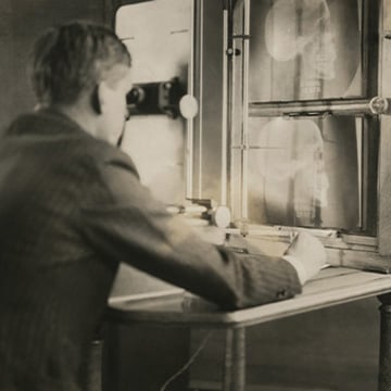
Photo: Cleveland Clinic Archives
THEN
A Cleveland Clinic radiologist analyzes a head X-ray in the 1920s. EARLY MEDICAL IMAGING included X-rays, discovered in 1895, and ultrasound, first used for medicine in the 1940s. It wasn’t until the 1970s that computerized axial tomography (CAT), a device capable of directly imaging pathology of the brain in a cross-sectional display, was developed. Realizing its potential, Chair of Radiology Thomas F. Meaney, MD, purchased the fourth such device in the world for Cleveland Clinic. In his honor, the Eastman Kodak Company established the Thomas F. Meaney, MD, Research Scholars Endowed Chair; today, it’s held by Imaging Institute Chair Greg Borkowski, MD, FACR. In the early 1980s, Cleveland Clinic’s Department of Diagnostic Radiology led the way in developing nuclear magnetic resonance imaging, later called magnetic resonance imaging (MRI). Instead of using X-rays to produce images like a CT scan, MRI uses a large magnet, radio waves and a computer to produce detailed images.
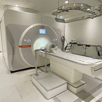
Photo: Annie O'Neill
NOW
Cleveland Clinic is one of the few institutions worldwide with a 7-TESLA (7T) MRI SCANNER. MRI scanners come in different magnet field strengths, measured in tesla units. The higher the tesla, the greater the image resolution and tissue contrast. Cleveland Clinic built a dedicated 2,300-square-foot research facility to house the machine with its 80,000-pound magnet. This advanced imaging technology, approved for clinical use by the FDA in 2017 but used for research at Cleveland Clinic since 2014, has had a notable impact on the diagnosis and treatment of multiple sclerosis, epilepsy, traumatic brain injury, degenerative brain disorders and brain tumors.
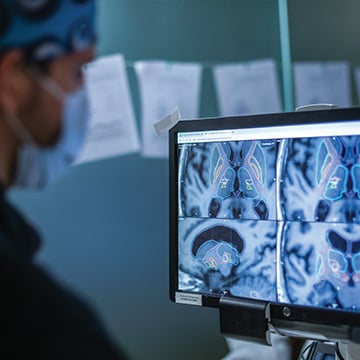
Photo: Annie O'Neill
NEXT
A team led by Cleveland Clinic neurosurgeon Sean Nagel, MD, uses HIGH INTENSITY FOCUSED ULTRASOUND (HIFU) to pinpoint treatment for a patient with Parkinson’s disease. Similar to how a magnifying glass focuses light on a target, HIFU produces multiple beams of ultrasound concentrated on the exact tissue area requiring treatment and uses the energy to destroy the targeted area. The ultrasound beams are able to pass through layers of tissue, leaving them unharmed, until they reach their target — essentially, surgery without a scalpel. Patients with debilitating tremor are already being successfully treated by precisely targeting the spot in the brain causing the tremor with HIFU, but researchers are excited about its broader potential. “We’re investigating other brain diseases that may benefit from HIFU treatment, such as major depression and epilepsy,” says Stephen Jones, MD, PhD, Vice Chair of Research and Academic Affairs in Cleveland Clinic’s Imaging Institute. “Researchers from our Imaging Institute and Neurological Institute also are exploring other new ways to precisely image potential targets — using 7T MRI, for example, to help treat disease without an incision.”